Introductory price — today only

Digital book in PDF: it comes by email. Nothing is shipped on paper.

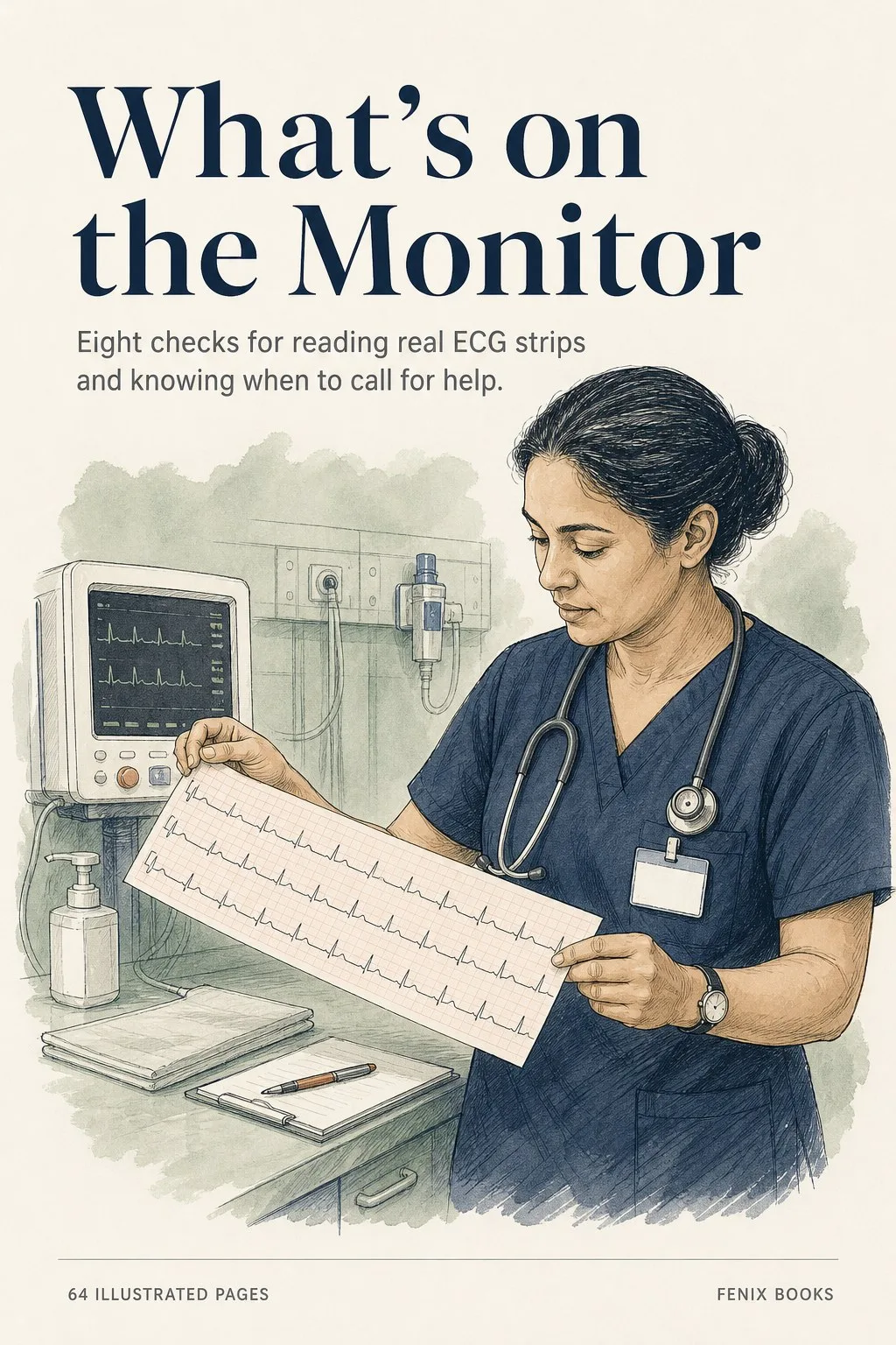
The ECG strip guide that starts with the patient and walks you through eight checks to a decision

Practice measuring intervals, describing a rhythm, and organizing an SBAR report with annotated examples and five practice strips.

Get the guide ➔

Protected checkout · PDF by email · 30-day guarantee

68pages total, including 64 topic pages
8sections you can jump between
2free bonus books included
PDFby email, printable

Read the measurements on these sample pages

Made to be read on your phone during a break or between shifts, or printed for your binder.

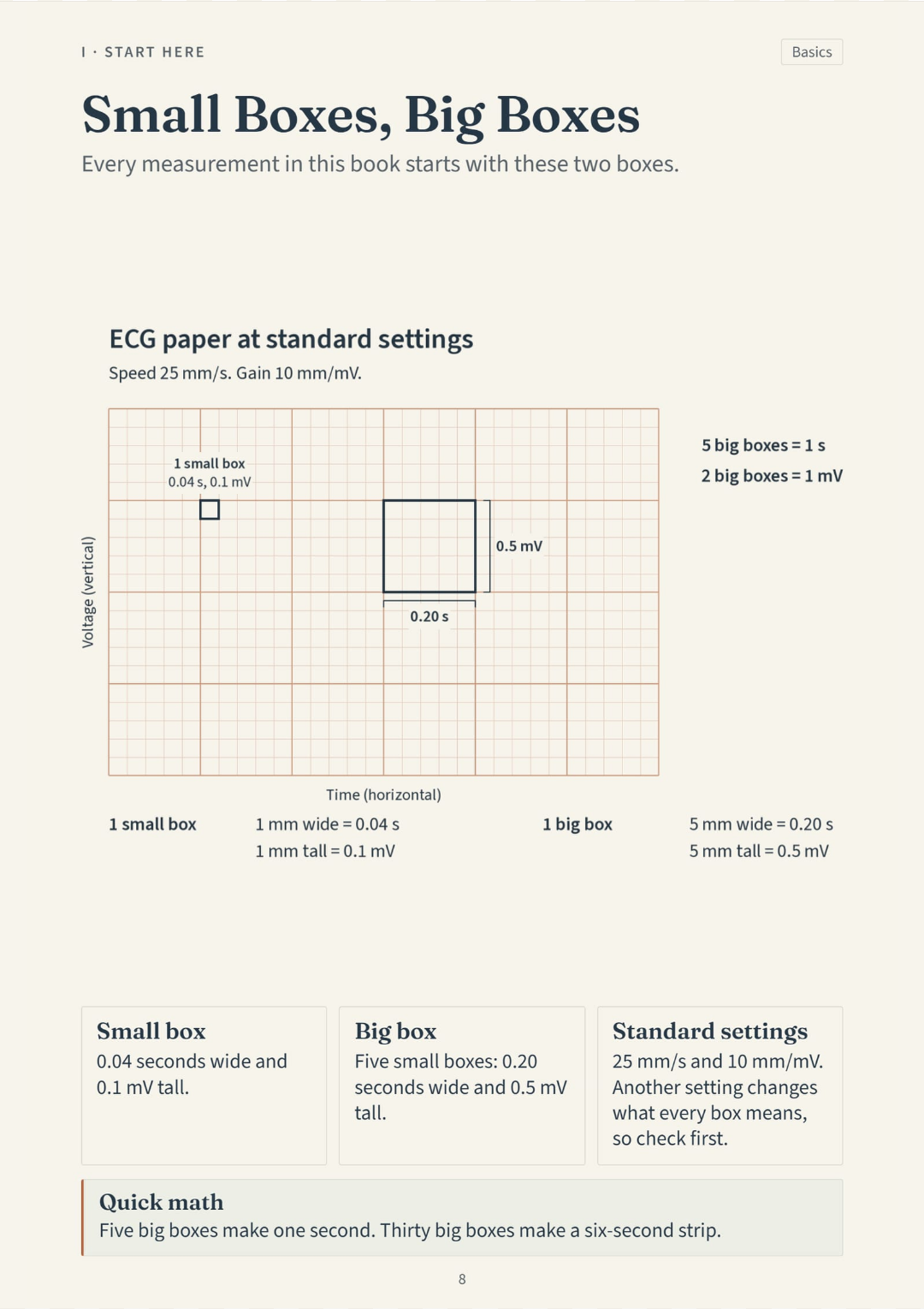
Every measurement in this book starts with these two boxes. One small box is 0.04 seconds; one big box is 0.20 seconds.
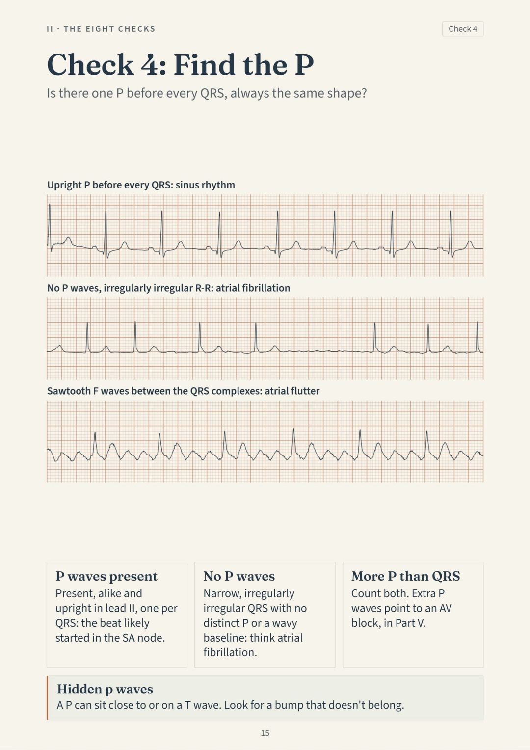
Is there one P before every QRS, always the same shape? P waves tell you where the beat started and how it traveled.
Irregularly irregular, with no P wave you can point to. No P waves, no pattern in the R-R: atrial fibrillation.
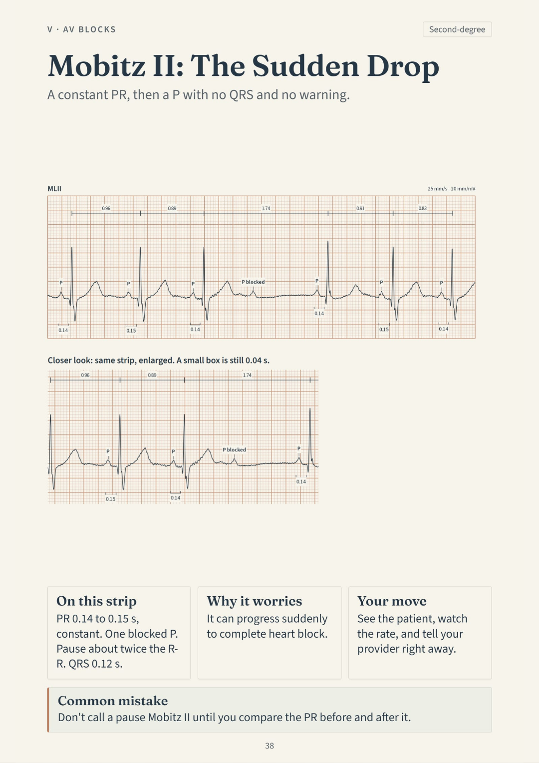
A constant PR, then a P with no QRS and no warning. Constant PR with a sudden blocked P: Mobitz II, report it right away.
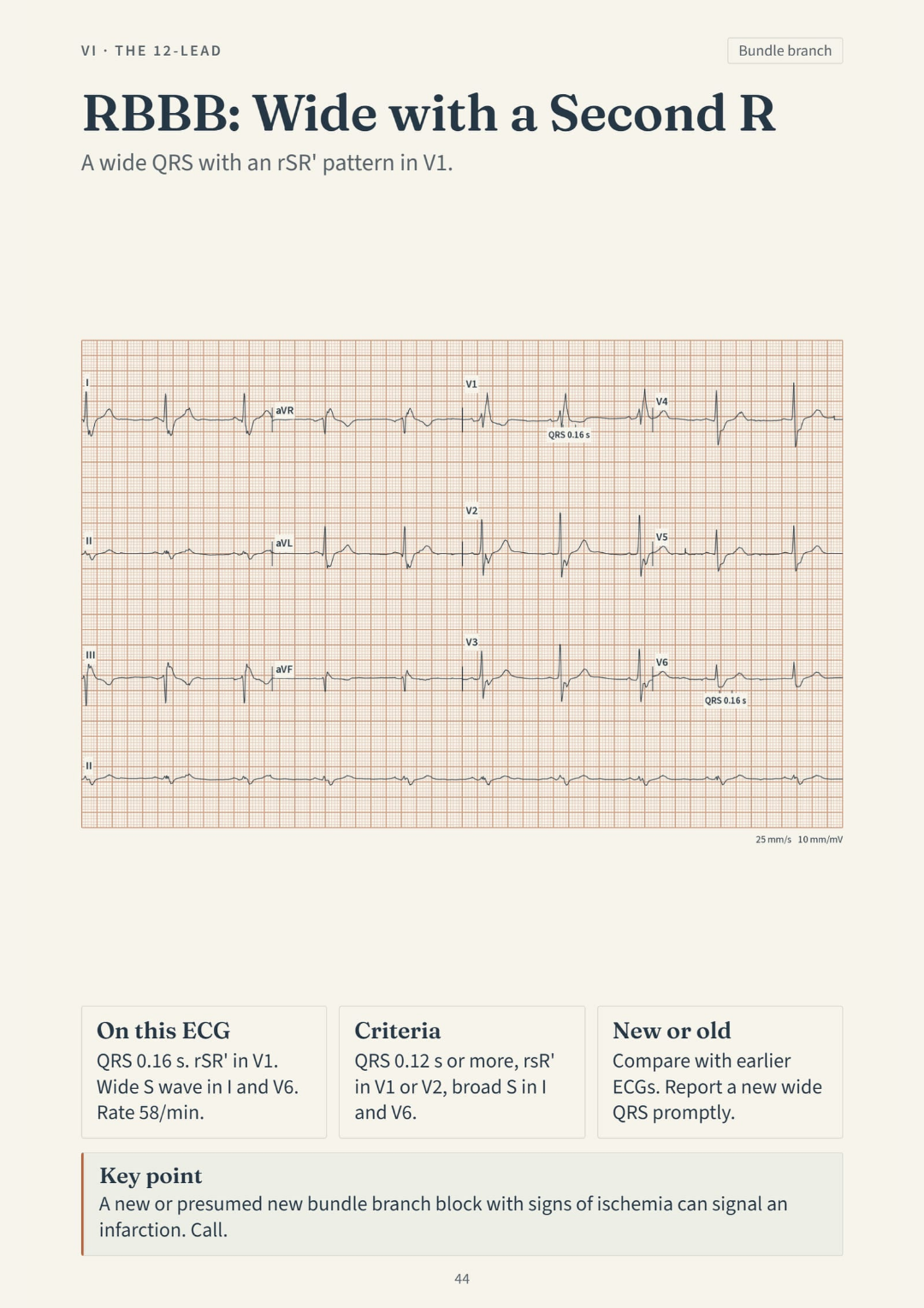
A wide QRS with an rSR' pattern in V1. Wide QRS plus a second R in V1: right bundle branch block.
ST up in II, III, aVF. ST down in I, aVL. ST up in the inferior leads, mirrored down in I and aVL.
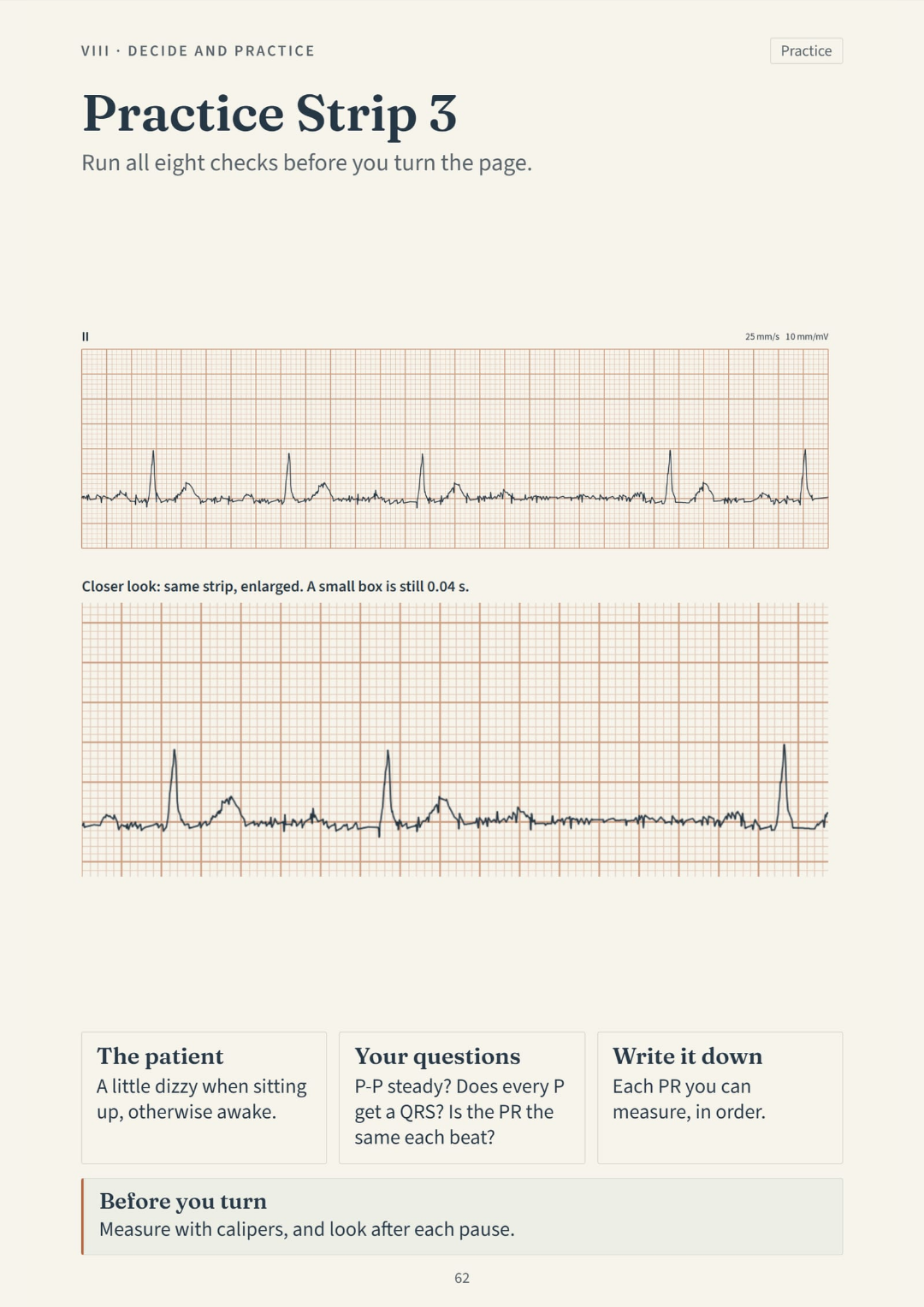
Run all eight checks before you turn the page. An unlabeled tracing: run the eight checks, then turn the page.

Swipe to see more · tap to zoom →

Naming a rhythm is only part of the report. What observations support it?

You may recognize a rhythm name yet hesitate when asked to explain it. Which intervals did you measure? Are the R-R intervals regular? What did you observe about the P waves?

During a study session, an uncertain P wave can derail your explanation. Write down the feature you cannot resolve, then compare it with the annotated example or ask your instructor.

The book connects measurement with communication. Its rhythm pages lead into practice cases and an SBAR section, so you can rehearse both the observations and the report.

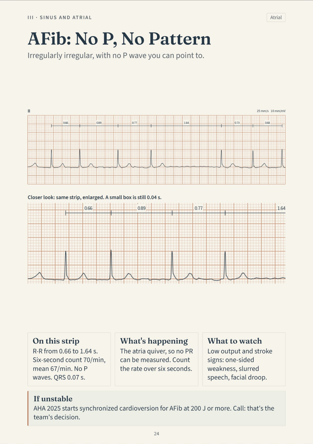
Irregularly irregular, with no P wave you can point to. No P waves, no pattern in the R-R: atrial fibrillation.

No P waves anywhere. No pattern in the R-R intervals. That is atrial fibrillation, and the page shows you exactly what to look for — drawn on the tracing, with the numbers printed.

Use the AFib example to rehearse a specific description: the P waves and R-R intervals are the features to discuss.

A useful practice answer explains the observations behind the label.
COMPARE

Mobitz II: connect the definition to the measured intervals

INSTEAD OF THIS
  • Read a dense paragraph describing Mobitz II with abbreviations, exceptions, and cross-references.
  • Try to work out what a dropped beat looks like from the wording alone.
  • Hope the difference between Mobitz I and Mobitz II sticks when the alarm goes off.
Second-degree atrioventricular block, Mobitz type II, is characterized by intermittent failure of atrial impulses to conduct to the ventricles without progressive prolongation of the PR interval. The PR of conducted beats remains constant, distinguishing it from Mobitz type I (Wenckebach), in which the PR progressively lengthens before the nonconducted beat. The block localizes to the infranodal conduction system and the conducted QRS may show a bundle branch block pattern. Mobitz II carries significant risk of progression to complete heart block and hemodynamic compromise, requiring continuous monitoring with transcutaneous pacing available and early consultation for possible permanent pacemaker placement.
CHOOSE THIS
  • See the constant PR and the sudden dropped beat drawn right on the tracing.
  • The measured intervals are printed right on the strip, in the same place every time.
  • Every rhythm has its own spot in the book. Flip back to it at three in the morning if you need to.
A constant PR, then a P with no QRS and no warning. Constant PR with a sudden blocked P: Mobitz II, report it right away.
CONTENTS

Find the rhythm, measurement, or practice case you need

Eight sections cover the grid, rhythm checks, AV blocks, the 12-lead, and practice cases. The 68-page PDF contains 64 topic pages plus the cover and contents.

IStart Here · pp. 5-10
  1. Read This First
  2. Patient First, Monitor Second
  3. The Heart's Wiring
  4. Small Boxes, Big Boxes
  5. One Beat, Piece by Piece
  6. Tele Leads on the Chest
IIThe Eight Checks · pp. 11-19
  1. The Eight Checks, in This Order
  2. Check 1: Patient and Signal
  3. Check 2: Rate
  4. Check 3: Regular or Not
  5. Check 4: Find the P
  6. Check 5: The PR Interval
  7. Check 6: QRS Width
  8. Check 7: QT and ST-T
  9. Check 8: Decide and Tell
IIISinus and Atrial Rhythms · pp. 20-27
  1. Normal Sinus: All Eight Checks
  2. Sinus Bradycardia
  3. Sinus Tachycardia
  4. PAC: The Early Narrow Beat
  5. AFib: No P, No Pattern
  6. Flutter With a Fixed Ratio
  7. Flutter 2:1: The Steady 150
  8. SVT: Narrow, Fast, Regular
IVVentricular Rhythms and Arrest · pp. 28-34
  1. PVC: Early, Wide, No P
  2. Paced Rhythm: Spike, Then QRS
  3. VT: Wide, Fast, Same Shape
  4. Torsades: The Twisting Rhythm
  5. VF: Chaos and No QRS
  6. Asystole and PEA
  7. No Pulse: What You Do
VAV Blocks · pp. 35-40
  1. Four Blocks, One Ladder
  2. First-Degree: Late Every Time
  3. Mobitz I: The Stretching PR
  4. Mobitz II: The Sudden Drop
  5. High-Grade Block: 3 P, 1 QRS
  6. Third-Degree: P and QRS Apart
VIThe 12-Lead · pp. 41-47
  1. Getting a Clean Tracing
  2. How 12 Leads Are Laid Out
  3. Axis: Start With Two Leads
  4. RBBB: Wide With a Second R
  5. LBBB: Broad R on the Left
  6. WPW: The Delta Wave
  7. Tall Voltage: The LVH Pattern
VIIIschemia and Infarction · pp. 48-54
  1. Which Leads See Which Wall
  2. What Counts as ST Elevation
  3. ST Elevation in V2 to V6
  4. Inferior, With a Mirror Image
  5. ST Depression in V5 and V6
  6. When ST Elevation Isn't a Blockage
  7. Chest Pain on Your Shift
VIIIDecide and Practice · pp. 55-68
  1. Code, Rapid Response or Provider
  2. SBAR: Say It in Four Parts
  3. The Urgent Sheet
  4. Practice Strip 1
  5. Practice Strip 1: Answer
  6. Practice Strip 2
  7. Practice Strip 2: Answer
  8. Practice Strip 3
  9. Practice Strip 3: Answer
  10. Practice Strip 4
  11. Practice Strip 4: Answer
  12. Practice Strip 5
  13. Practice Strip 5: Answer
  14. Credits and Sources

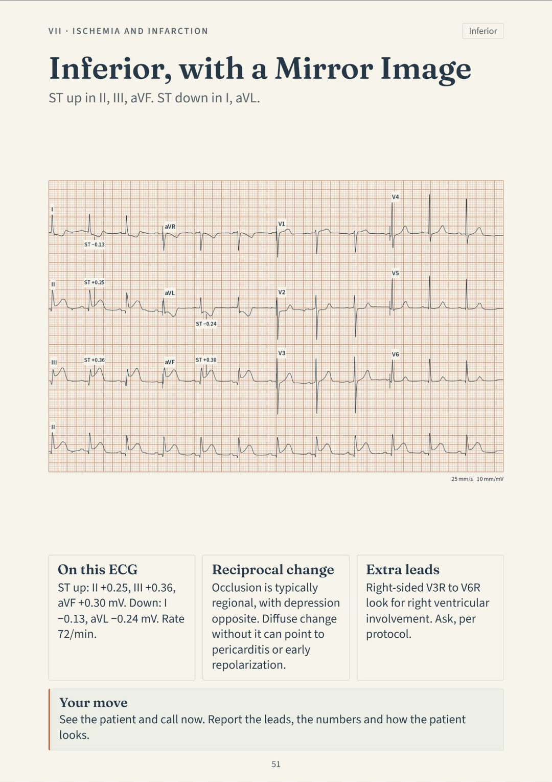
An inferior-lead example, with the changes labeled

Each topic pairs a concise explanation with a tracing, diagram, or checklist. Patient-recorded signals are replotted from research databases; simulated strips are labeled. Measurements and study notes help you connect the pattern to what you should report.

ST up in II, III, aVF. ST down in I, aVL. ST up in the inferior leads, mirrored down in I and aVL.

ST elevation in leads II, III, and aVF. ST depression in I and aVL — the mirror image. Both are on the same page, with the numbers marked.

Read the lead labels alongside the marked ST changes, then compare your description with the notes beneath the tracing.

The example links a location on the tracing with a finding you can describe.

Study sections, from the grid to the handoff

Ipp. 5-10

Start Here

The grid, the wiring, and how tele leads sit on the chest.

IIpp. 11-19

The Eight Checks

The eight checks in order: patient, rate, regularity, P waves, PR, QRS, QT and ST-T, then decide.

IIIpp. 20-27

Sinus and Atrial Rhythms

Sinus rhythms, PACs, AFib, flutter, and SVT — each on its own page.

IVpp. 28-34

Ventricular Rhythms and Arrest

PVCs, paced rhythm, VT, torsades, VF, and asystole — and how to recognize when you need to check for a pulse and call for help.

Vpp. 35-40

AV Blocks

Four AV blocks on one ladder: first-degree through third-degree.

VIpp. 41-47

The 12-Lead

The 12-lead: placement, axis, bundle branch blocks, WPW, and LVH.

VIIpp. 48-54

Ischemia and Infarction

Ischemia and infarction: which leads see which wall, and what counts as ST elevation.

VIIIpp. 55-68

Decide and Practice

When to call a code, a rapid response, or the provider — plus five practice strips with answers.

Two included references for medication and lab study

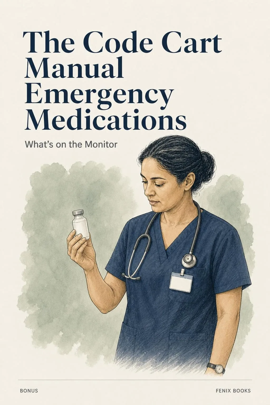
The Code Cart Manual: Emergency Medications

GIFT 1: «The Code Cart Manual: Emergency Medications»

Bought separately: US$ 22.90

Look up the adult emergency drugs by rhythm, all given only on an order or protocol: when each is used, the dose and what to watch. Written only from the AHA 2025 Part 9 guideline and its algorithms, the 2010 AHA/ACCF torsades statement, FDA labels in DailyMed and MedlinePlus drug information, with your hospital protocol always first.

Behind the Strip: The Lab Values Manual

GIFT 2: «Behind the Strip: The Lab Values Manual»

Bought separately: US$ 18.90

See which lab results can change the strip, which ones matter around it and what to report. Ranges from the ABIM Laboratory Reference Ranges, plain explanations from MedlinePlus, and your own lab's range always comes first.

Your bundle: What's on the Monitor and 2 gifts

  • What's on the Monitor: 64 illustrated pagesUS$ 74.90
  • GIFT 1: The Code Cart Manual: Emergency MedicationsUS$ 22.90
  • GIFT 2: Behind the Strip: The Lab Values ManualUS$ 18.90
  • High-quality PDF file, ready to printIncluded
Total value US$ 116.70
BUNDLE PRICE
US$ 29.90
YOU SAVE: US$ 86.80
GET WHAT'S ON THE MONITOR · US$ 29.90

Digital book in PDF: it comes by email. Nothing is shipped on paper.

Protected paymentEncrypted data30-day guarantee

PDF by email: Your purchase covers the guide and both companion books in PDF format. Enter the email address you want us to use for delivery. This is a single payment, with no installments or additional charges.

Choose the study task that matches your next rotation

New grad heading to a telemetry or stepdown unit

New grad heading to a telemetry or stepdown unit

The monitor is the part that scares you most. Practice a fixed order of eight checks, from the patient to the decision, with tracings replotted from research data and clearly labeled simulations.

Nursing student with a strip exam or cardiac rotation coming up

Nursing student with a strip exam or cardiac rotation coming up

You need practice describing a strip. Work through five unlabeled practice strips, each followed by an answer page, and review rhythm patterns with measured intervals.

Nurse preparing for ACLS or moving to a cardiac floor

Nurse preparing for ACLS or moving to a cardiac floor

You've done med-surg. Now you need to read rhythms every shift. This is the refresher built on AHA 2025 and the Fifth Universal Definition of MI, with sources named on every page. It doesn't replace your ACLS course.

The guide and companions in this order

US$ 116.70US$ 29.90
GET WHAT'S ON THE MONITOR · US$ 29.90

Digital book in PDF: it comes by email. Nothing is shipped on paper.

30DAYS
GUARANTEE

Your 30-day refund option

A full refund is available during the first 30 days if the materials differ from this description or do not help with the study questions you purchased them for. In your email, explain the mismatch or the question that remained unresolved.

Common questions

Is this a physical book or a digital one?

Digital. You receive a PDF by email. You can print it if you want a binder copy.

Can I read it on my phone?

Yes. Every rhythm is drawn to fit a phone screen, with its numbers on the strip.

Which sections should a new learner read first?

Start with Part I for the grid, cardiac wiring, and lead placement. You can begin without prior ECG knowledge, then use the later practice pages to review those foundations.

Where do I get the book?

You get it as a PDF by email, sent to the address you enter at checkout.

Will I be charged again later?

No. There's one charge, and the PDF is yours to keep.

Is this an official AHA or ACLS product?

No. This is an independent study guide for reading rhythms. It doesn't come with a certificate or CE hours, and it doesn't replace an ACLS course. It's written for US practice. Sources — including AHA 2025 and the Fifth Universal Definition of MI — are named on every page. Not affiliated with or endorsed by AHA, ACC, or any other organization.

Does this replace my nursing program or hospital protocol?

No. This is study material. Your hospital protocol always comes first, and the book says so on the first page.

Are the tracings from real patients?

They are not photos of monitor strips. The tracings are redrawn from public research databases, and any simulated tracing is marked as simulated. None come from identified patients. Each one has its measured numbers printed on it.

I already have a textbook and YouTube is free. What does this add?

Use the five unlabeled practice strips to write your observations before viewing the answers. The annotated rhythm pages and SBAR section let you revisit a measurement or rehearse how you would communicate it.

Pick a strip for your next study session

Practice describing the strip before someone asks you to do it on the unit.

Work through the checks, then compare your answer with the study notes.

Keep your clinical training and local protocol first.

Bring a tracing to your next study session. Work through it in order, compare the answer, and take the questions you still have to your instructor.

US$ 29.90, paid once · 30-day guarantee · PDF by email

GET WHAT'S ON THE MONITOR · US$ 29.90

Digital book in PDF: it comes by email. Nothing is shipped on paper.